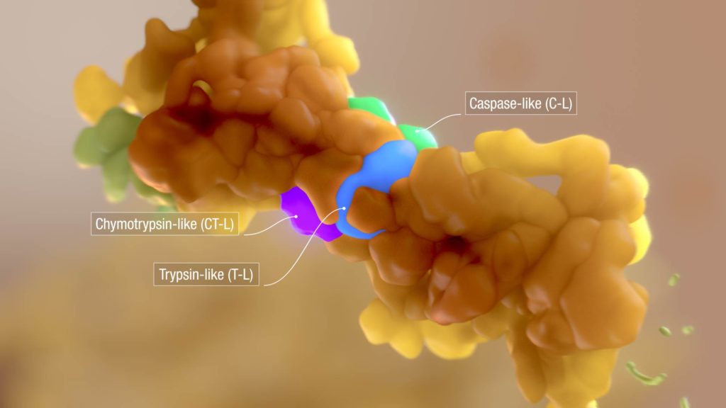

Detailed 3D rendering of the proteasome, a key macromolecular machine regulating cell health by degrading unwanted proteins. In this image, the enzymatic subunits of the proteasome are labelled: Chymotrypin-like (CT-L); Caspase-like (C-L); Trypsin-like (T-L). From a medical animation about the function of the proteasome in unwanted protein degradation. Myeloma cells exploit this function to survive under conditions of ER stress, via the unfolded protein response (UPR). © 2016. All Rights Reserved.
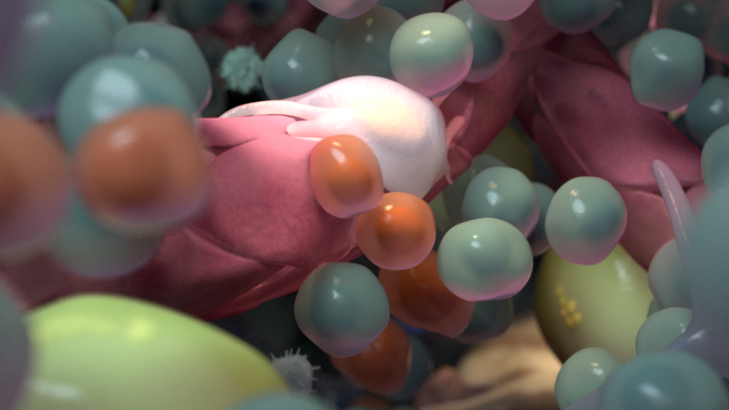
ER stress. The accumulation of misfolded proteins in the endoplasmic reticulum results in ER stress that can lead eventually to cell death. From a medical animation about the function of the proteasome in unwanted protein degradation. Myeloma cells exploit this function to survive under conditions of ER stress, via the unfolded protein response (UPR). © 2016. All Rights Reserved.
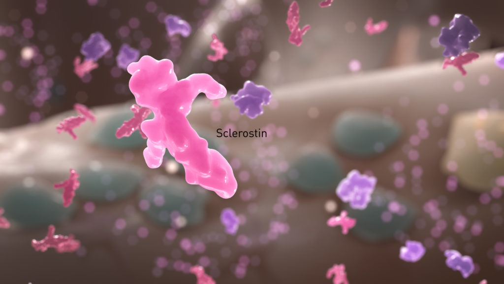
Lytic bone lesions in multiple myeloma. From a medical animation about the function of the proteasome in unwanted protein degradation. Myeloma cells exploit this function to survive under conditions of ER stress, via the unfolded protein response (UPR). © 2016. All Rights Reserved.