
Medical Animation: CGRP in Migraine
Created for Biopharmaceutical Client
Link copied!

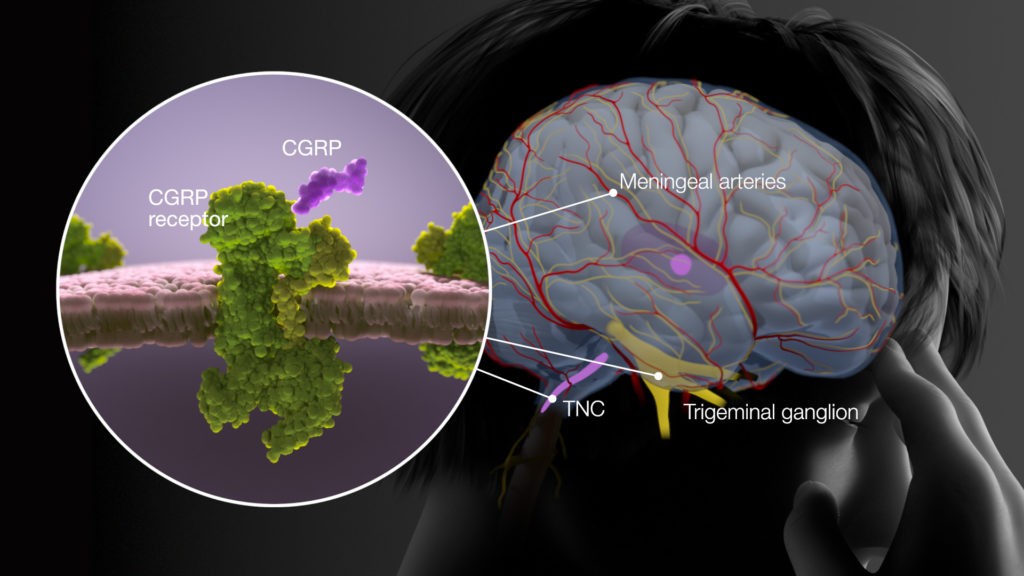
Locations in the brain (meningeal arteries, TNC, and trigeminal ganglion) where CGRP binds to CGRP receptors during a mirgaine. © 2017. All Rights Reserved.

Profile view of a female patient with brain anatomy overlayed, defining the cortex, thalamus, trigeminal nerve fibers, trgeminal nucleus caudalis (TNC) and trigeminal ganglion. © 2017. All Rights Reserved.

Synaptic cleft showing the release of CGRP from the presynaptic cleft to the postsynaptic cleft where the CGRP receptors are located. © 2017. All Rights Reserved.

Profile view of a female patient with brain anatomy overlayed (nerve and vascular network) © 2017. All Rights Reserved.
The project
We created this medical animation to educate headache specialists on the latest information about the hormone CGRP in migraine pathophysiology. Beginning with an infographic approach to burden of disease, the story moves to common migraine symptoms and then CGRP and CGRP receptor localization the CNS and PNS. We used a limited color palette and tight camera framing to emphasize the emotional aspects of migraine pain. Our voiceover script was based on a thorough review of the migraine literature, while the anatomy and molecular visuals were informed by data, including peer reviewed literature.
Created for
Biopharmaceutical client