1. Realistic Human Anatomy from Scan Data
The innovation: Realistic human anatomy models are generated from CT or MRI scans and cleaned up for use in the 3D medical animation workflow.
Why it matters: Anatomical models can be created more quickly and accurately than with standard manual 3D modeling techniques. This saves time and helps ensure the high level of accuracy our biopharma and medical device clients require in their animations.
The DICOM image stack on the right contains scan data for all body tissues. The 3D skeleton on the left has been segmented from the scan data using advanced image processing technology . The resulting 3D model is highly accurate and saves hours of manual 3D modeling.
DICOM image processing applications like Osirix make this workflow possible. Previously, medical animators created models manually, pushing and pulling mesh vertices with digital sculpting tools while consulting 2D references of the structure they were modeling. Now, much of that process is automated, but still requires some artistry and skill. To start, the animator uses the image processor to segment, or isolate, the anatomy they need. The resulting model is a 3D surface mesh that typically requires digital clean up and optimization before it can be used in a 3D animation program.
With the number of anonymized DICOM scans in public databases increasing all the time, it’s possible to find and model a wide range of anatomical variation and pathologies.
2. Data-Driven Molecular Visualization
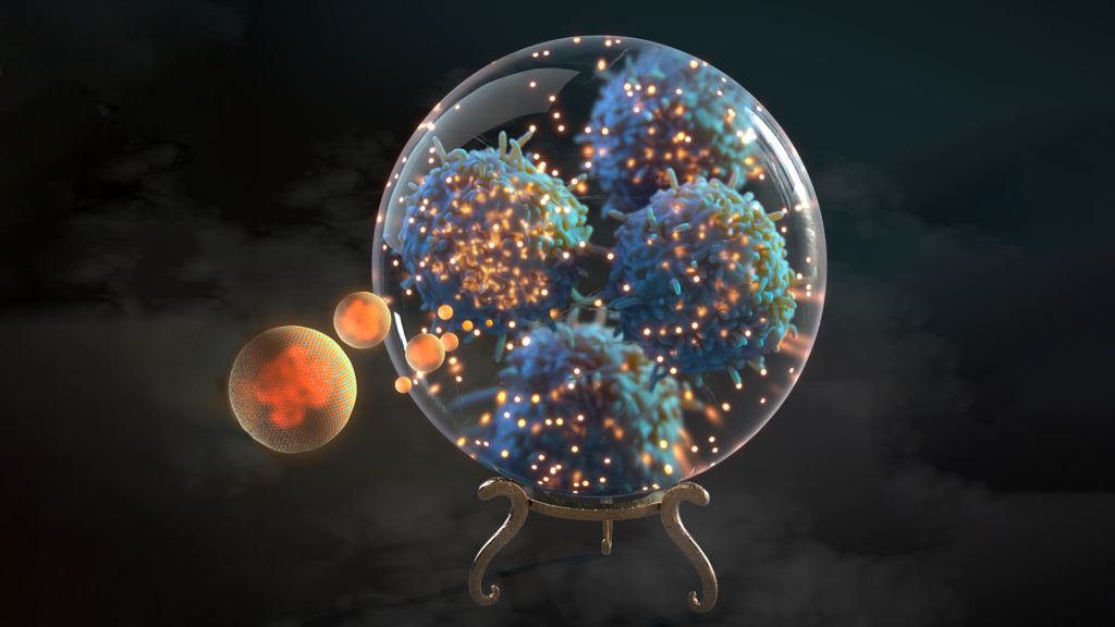
The innovation: The spatial coordinates of every atom in a complex biomolecule such as DNA or a protein are obtained using x-ray crystallography or nuclear magnetic resonance (NMR). Scientists make these data available through public datasets, where animators can use them to generate 3D models with unprecedented detail for use in medical animations.
Why it matters: The size and shape of biomolecules are closely linked to their function. Medical animators can depict molecules at accurate scales and show audiences how they interact with one another. This is important for teaching molecular biology, explaining disease and drug mechanisms, and research.
With access to molecular structure data, medical and scientific animators can visualize molecular activity at an unprecedented level of spatial detail.
A great many biomolecules can be modeled from structural data. For molecules whose structures are not yet mapped, medical animators may use AlphaFold, an artificial intelligence (AI) model that predicts structure with a level of accuracy sufficient for most medical animations. Nonetheless, while much of the modeling process can be automated using custom computer scripts or plugins, medical animators still make many artistic decisions related to the level of detail shown, motion and cinematography.
3. VR and AR Integration
The innovation: Virtual reality (VR) and augmented reality (AR) technologies enable immersive medical animations that can incorporate interactivity.
Why it matters: VR allows audiences to experience scientific content while immersed in the action, making learning more impactful and memorable. AR blends the real and digital worlds to enable richer learning experiences.
Rethink Osteoporosis integrates AR with medical animation to teach physicians about fragility fractures in an engaging way.
With VR, AR and mixed reality (MR), we’re able to make learners part of the medical animation story. See our MiBrain and PsA Simulator for examples. Actively participating in the story makes the content more relatable than passively watching or reading about it.
4. Interactive Medical Animations
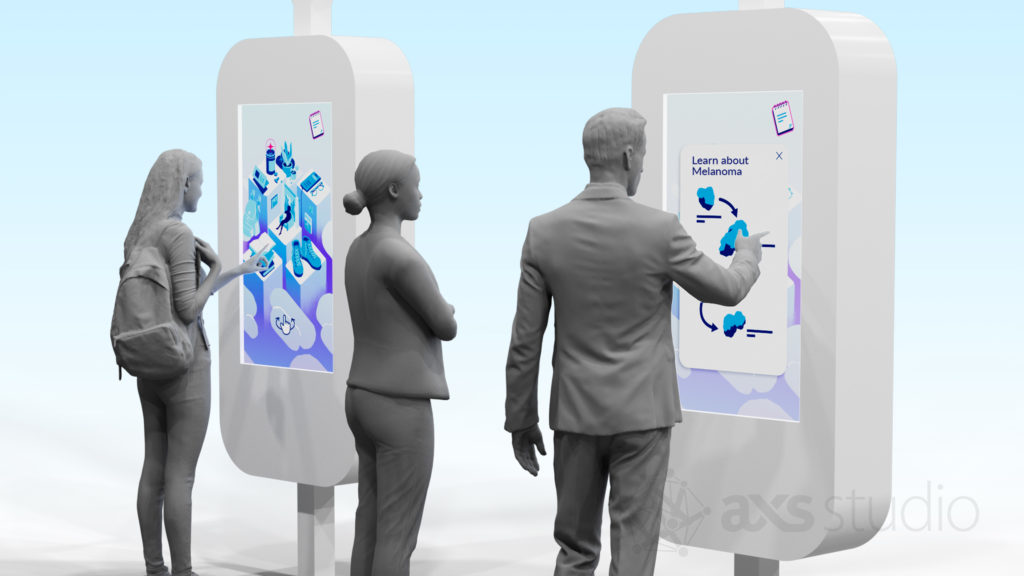
The innovation: Interactive elements are incorporated into the video, allowing viewers to make choices and have agency in the narrative.
Why it matters: Interactivity encourages user engagement and improves retention of the material. Interactivity in medical animation enhances clinician and patient education, and makes marketing videos more effective.
Interactive medical animation ranges from linear narratives with clickable elements to fully gamified immersive non-linear experiences. As GPU rendering capabilities advance, AXS Studio is increasingly using game engines to develop real-time interactive animations that audiences can explore at their own pace.
5. Storytelling for Patient Education
The innovation: Medical animations integrate narrative storytelling and engaging visuals to simplify complex topics for patients and caregivers.
Why it matters: When patients understand their condition and treatment options, they can feel more empowered and make informed decisions about their care.

Aimed at Alzheimer’s disease (AD) patients and caregivers, this medical animation depicts a cerebrospinal fluid donation procedure in support of an AD research project. It uses plain language, relatable characters and simplified depictions of anatomy and molecules.
Disease pathophysiology and therapeutic interventions are complex and often difficult for those without medical training to understand. Physicians often lack sufficient resources to educate their patients on topics with potentially life-altering implications. To address this issue, medical animators with graduate training in medical science and filmmaking have optimized short-format film narrative to explain topics clearly and concisely—usually in 3 minutes or less. These engaging medical animations use plain language and can be easily shared with caregivers, family and friends.
6. Procedural 3D Scene Generation
The innovation: Complex organic 3D models are generated automatically using rules set up by the animator. These models can be quickly modified and refined by tweaking the parameters of these rules.
Why it matters: Micro- and nanoscopic tissue environments are common features of medical animations. Creating these scenes using manual 3D software tools can be difficult and time-consuming to modify. Procedurally generated models are inherently flexible, making medical animators more responsive to changes in direction.
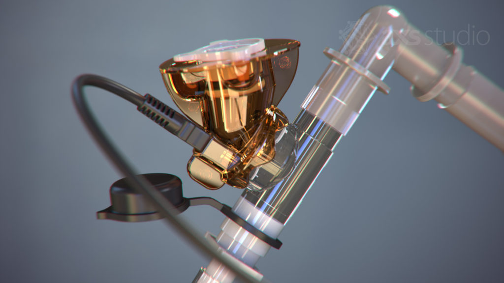
This model of a fascicle of sensory nerve axons was modeled entirely procedurally. The number of axons, lengths of the myelinated sections, widths of the nodes of Ranvier and other properties were controlled by numeric parameters. These were easily changed in response to feedback from the Medical Director, as we worked together to balance visual clarity and scientific accuracy. The finished medical animation can be seen here.
Procedural techniques have dramatically changed 3D modeling. They enable artists to add and modify organic details in anatomical, cellular and molecular scenes with unprecedented flexibility. Custom procedural workflows allow rapid experimentation to create visually compelling medical animations, while being responsive to changes as project requirements evolve.
7. Retopology of 3D Geometry
The innovation: Retopology is the process of optimizing the polygonal mesh of a 3D model to reduce its complexity for more efficient rigging, animation and rendering of medical animations.
Why it matters: Anatomical models generated from CT or MRI data (see above) have a lot of detail, meaning they are composed of many small polygons. This makes them difficult and slow to work with in the animation workflow. Retopology greatly reduces the number of polygons, while maintaining surface detail. The retopologized models are much “lighter” to work with, allowing medical animators to work more quickly and efficiently, without sacrificing important anatomical detail.
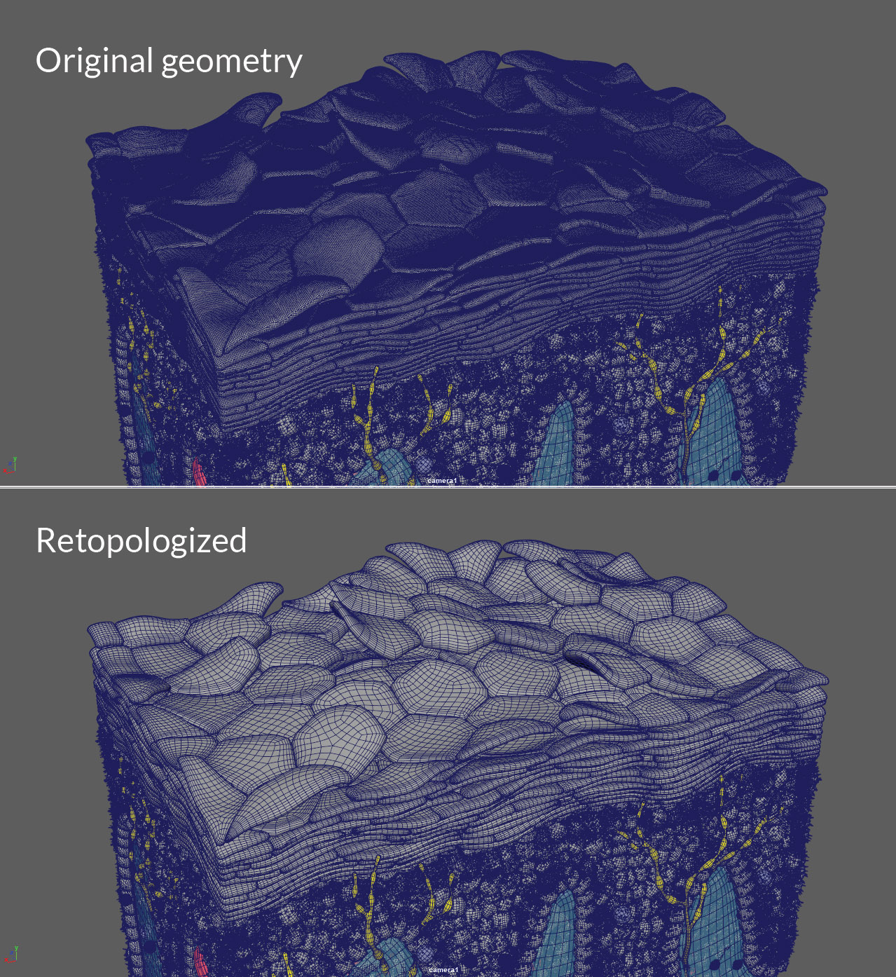
The stratum corneum of this human skin model was modeled procedurally, then digitally sculpted, resulting in a very dense, complex polygonal mesh. Retopology greatly reduced the number of polygons while retaining the shape of each corneocyte. This saved considerable time when we rendered this scene for the animation.
Retopology is used for more than anatomical models from scan data. Models that are manually built in 3D software benefit from retopology, as it evenly distributes polygons and unifies their size. The same applies to 3D models generated from medical device CAD files and models of human characters. Retopology tools have come a long way in recent years and are now available in most 3D animation and modeling software.
8. GPU Rendering
The innovation: Rendering 3D animations using a computer’s graphic processing unit (GPU) instead of its central processing unit (CPU) to dramatically reduce rendering times.
Why it matters: Rendering is the process of turning 3D scenes on a computer into finished videos and images. GPU rendering is many times faster and more cost effective than CPU rendering. As a result, medical animation studios can see finished results and test modifications much more quickly. The cost savings allow for increased production capacity. GPU rendering is also necessary for real-time immersive medical animation using VR or AR.
Because GPUs are optimized for parallel processing, they’re much faster for small, repetitive tasks like rendering compared to CPUs, which process information sequentially. GPUs can render in real- or near-real time, giving animators almost immediate feedback as they set up scenes in a medical animation. This makes the process of optimizing the finished look more efficient than with a traditional CPU rendering workflow. That said, for all of the advantages of GPU rendering, CPUs still provide an edge in rendering large, memory-intensive scenes and are therefore still utilized in animation production.
9. Cloud Render Farms
The innovation: Using powerful, unlimited cloud computing to render medical animations quickly, without requiring an on-site render farm.
Why it matters: Cloud farms can drastically accelerate the rendering process, while the pay-as-you-go model can save medical animation companies money and reduce the burden of technology upkeep.
Before the advent of cloud computing, animations had to be rendered on local networks of dedicated rendering computers, or farms. Animators were limited to the number of CPU and/or GPUs on hand, which in turn limited the speed and efficiency of production. Cloud farms make large numbers of high-performance “render nodes” available to anyone, without a capital cost investment. They also provide access to the latest hardware without the need for upgrades or maintenance. Cloud rendering is especially useful for tight deadlines, however the cost can be significant for larger projects, and the experience and reliability is entirely dependent on the third-party cloud farm company.
10. Essential Graphics
The innovation: Essential Graphics makes it easy to deploy consistent graphical elements—including motion graphics (mograph) and text—to multiple scenes in a medical animation at the editing and compositing stage.
Why it matters: Graphical elements are used through medical animations to augment the visuals. The Essential Graphics workflow leverages templates to add these repeated elements throughout and between animations. It saves time, maintains consistency, and makes global changes efficient.
The Essential Graphics workflow leverages templated motion graphics to maintain coherence and brand consistency across medical animations. See the full scientific animation here.
At AXS Studio, we build a design system to establish graphical and brand standards for the medical animations we create for each client. We use Essential Graphics to pull the design system into each production. The templates are shared between projects, resulting in a consistent look and feel throughout and across all of a clients’ animations.