You have dozens of organs that make your body work. But did you know that only 5 organs are considered essential? These fabulous five are your vital organs—the organs you absolutely cannot live without. What are the 5 vital organs of the body and what makes them the stars of the show?
The Brain
Why it’s a vital organ: The most bossy of the vital organs, the brain controls most essential processes in your body. It directly controls the functions of your other vital organs including your lungs (e.g., breathing), heart (e.g., heart rate), and kidney (e.g., glomerular filtration rate). Indirectly, your brain also controls certain functions of your vital organs. For example, hormones produced by your brain control fluid balance in the kidneys and metabolic processes in the liver.
Fun facts:
- Your brain was once on the outside of your body… well, sort of. In embryonic development, the tissue destined to become the brain—the neural plate—starts on the outside of the embryo. Through an intricate series of cell shape changes and movements, the neural plate rolls into a tube and moves inside the body. Once inside, the neural tube is patterned and folds to become the brain and spinal cord.
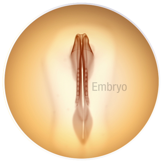
Chicken embryos, like the one in this video, develop in a very similar way as humans, making them an excellent model system for developmental biology research. As in humans, the tissue destined to become the chicken’s brain starts on the outside of the body. Intricate and coordinated cell shape changes create neural folds or small ridges on either side of the embryo’s midline. These folds fuse, rolling the neural plate into a tube shape and bringing the future brain inside the body.
- Your brain is harder to get into than Fort Knox. Being a vital organ, it’s no surprise the brain is heavily protected. Your brain is, of course, protected by your skull. Between your skull and your brain, the meninges and cerebrospinal fluid cushion your brain, protecting it from mechanical injury. Your brain is also protected by a highly selective “filter”, called the blood-brain barrier (BBB). Made up of endothelial cells, pericytes, and astrocytes, the BBB blocks harmful neurotoxins and pathogens in the bloodstream from entering and damaging the brain. While incredibly important for maintaining a healthy brain, the BBB poses a unique challenge when it comes to therapeutics. Drugs that need to access the brain must be specially designed to cross the BBB in order to work effectively.
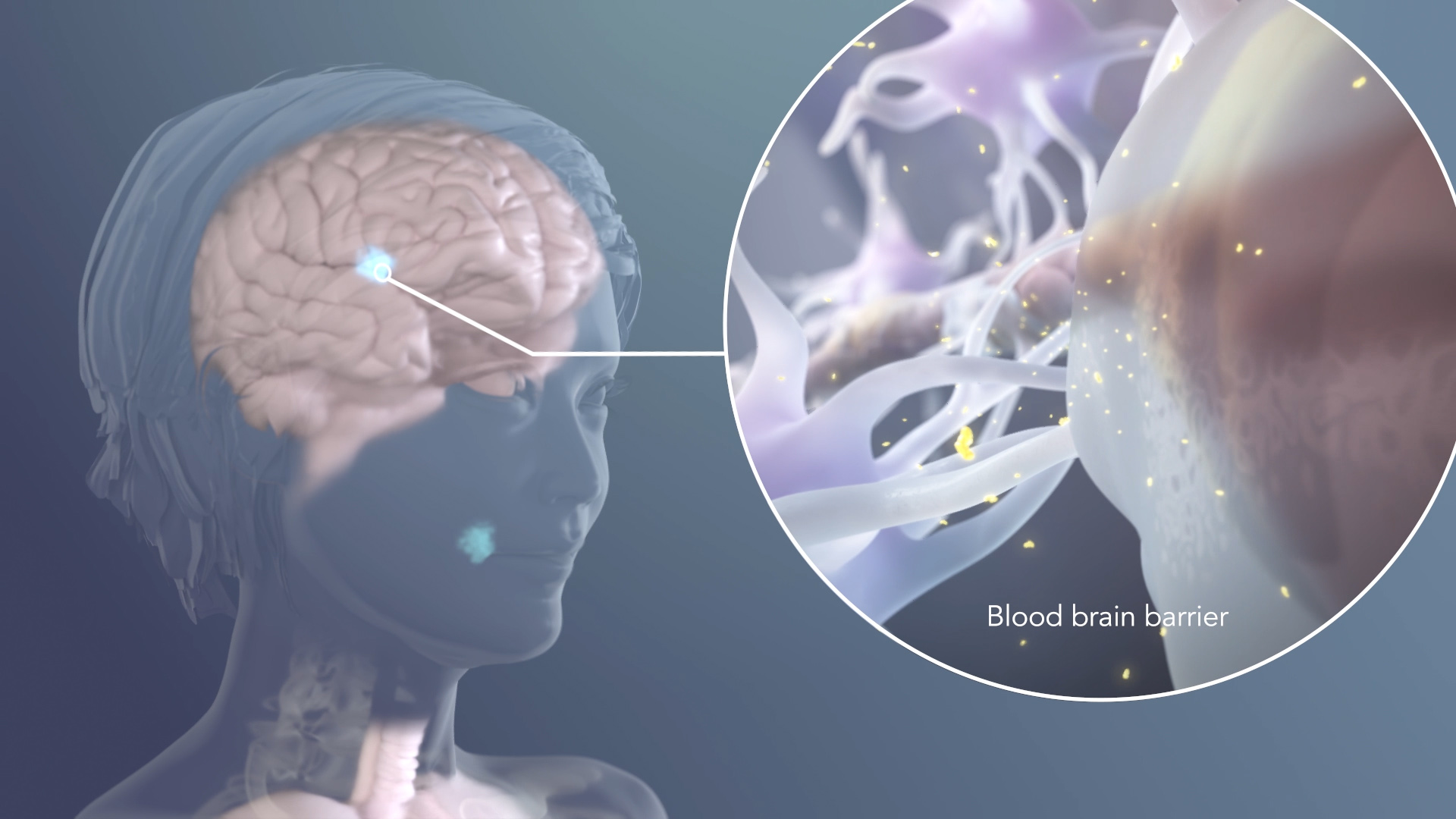
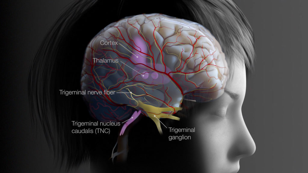
The blood-brain barrier (BBB) protects your brain from potentially harmful toxins and pathogens circulating in your blood. While indispensable for maintaining a healthy brain, the BBB is a bit of a double-edge sword. The BBB can also block the entry of helpful things, like therapeutics, from entering the brain. This image is from a mechanism of action animation about a small molecule drug used for the treatment of breast cancer metastases in the brain. The drug was specially engineered to cross the BBB so that it could access and treat brain metastases.
The Heart
Why it’s a vital organ: The centerpiece of your circulatory system, your heart is the vital organ responsible for pumping blood through the vast network of vessels within your body—60,000 miles (96,000 kilometers) of vessels, in fact.
Fun facts:
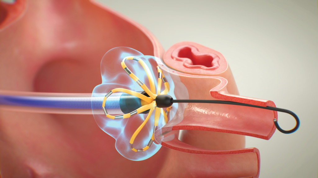
- Your heart has its own “brain”. Your heart has its own nervous system—the intrinsic cardiac nervous system (ICNS)—which precisely fine-tunes and coordinates your heartbeat. The ICNS allows your heart to quickly and independently adapt to varying conditions and demands, without having to rely on the brain. Disruptions to the ICNS can cause an abnormal heartbeat, or arrhythmia. In atrial fibrillation, electrical signals emanating from the left atrium abnormally trigger erratic heart contractions. Electrical signals from these malfunctioning cells can be blocked by a procedure called ablation in order to restore normal heart rhythm.
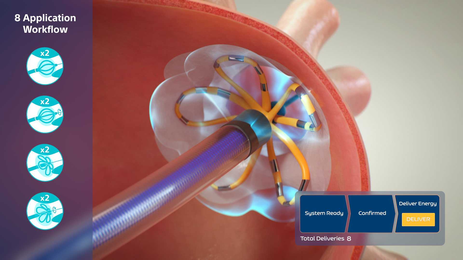
The medical device shown in this video is used to treat a heart arrhythmia called atrial fibrillation. A long catheter is fed into the heart, deploying the device inside the left atrium. The cardiac muscle cells responsible for the abnormal heart contractions are destroyed or ablated by an electrical field created by the device, while leaving surrounding cells and tissues unharmed.
- Heart contractions are driven by minute molecular motors. Cardiac muscle produces the force necessary to pump blood into circulation. Heart contractions visible at the gross anatomical level are driven by a tiny molecular motor protein called cardiac myosin. Using ATP as fuel, cardiac myosin cyclically binds and releases actin filaments to drive the shortening of sarcomeres, the basic contractile unit of heart muscle.
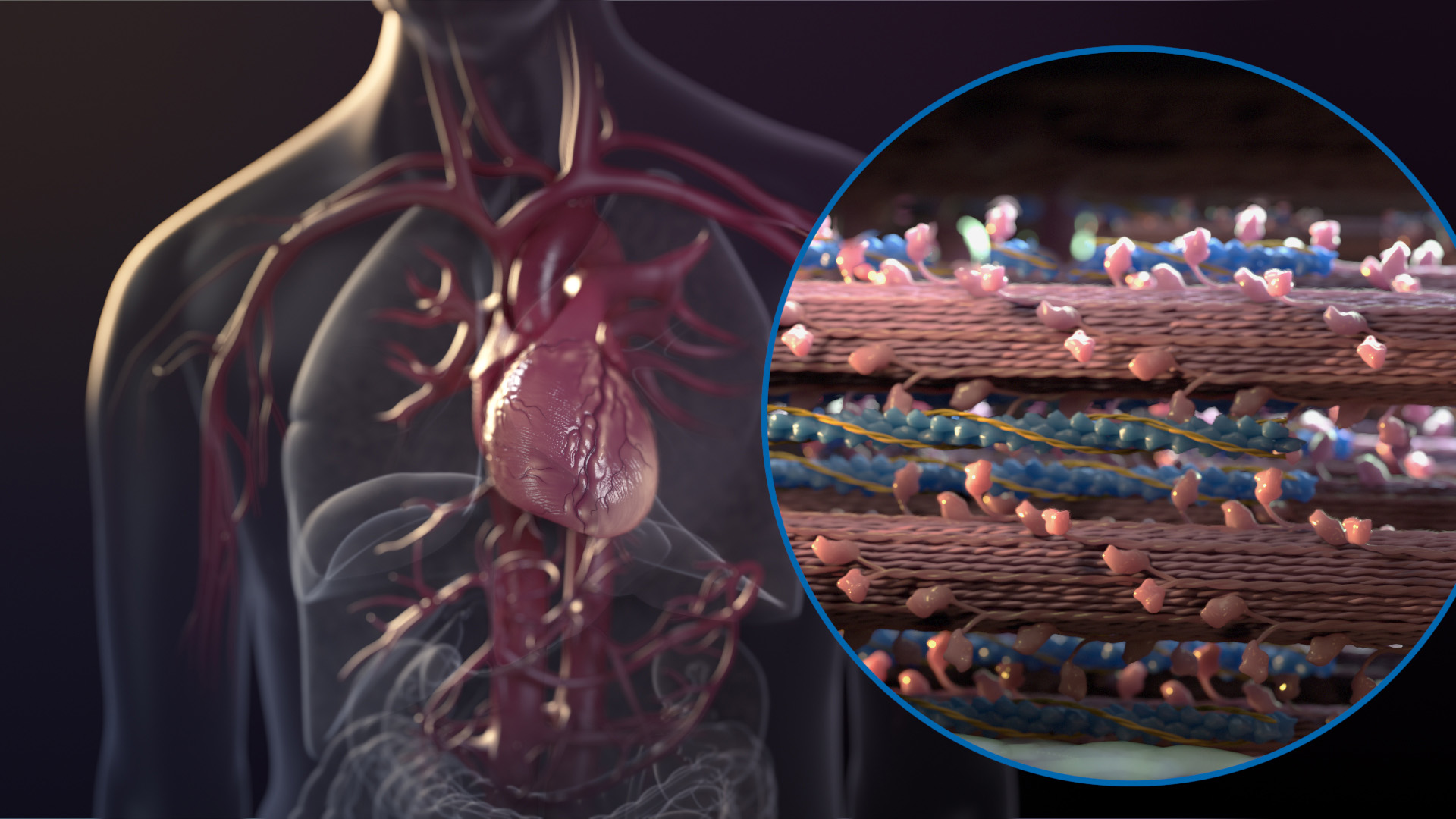
Heart muscle contractions start at the molecular level. In a coordinated and cyclical manner, cardiac myosin drives the shortening of sarcomeres, the basic contractile unit inside heart muscle cells. As shown in this video, this is a beautifully orchestrated process.
- Emotional heartbreak is real. Takotsubo cardiomyopathy—so called “broken heart syndrome”—is a temporary weakening of your heart muscle. Although its exact etiology is unclear, broken heart syndrome is thought to be triggered by stressful or emotional life events.
The Lungs
Why it’s a vital organ: The lungs are the vital organ responsible for gas exchange. Your lungs are not only the gateway for vital oxygen to enter your body, they also expel waste gas products, like carbon dioxide.
Fun facts:
- Your lungs are equipped with tiny brooms. The inside of your respiratory tract is lined with microscopic hair-like structures called cilia that sweep away mucus and debris from your lungs. They move in a coordinated, wave-like fashion. When this motion is impaired by thick mucus or by defects in the cilia themselves, such as in people with cystic fibrosis, lungs become susceptible to infection and damage.
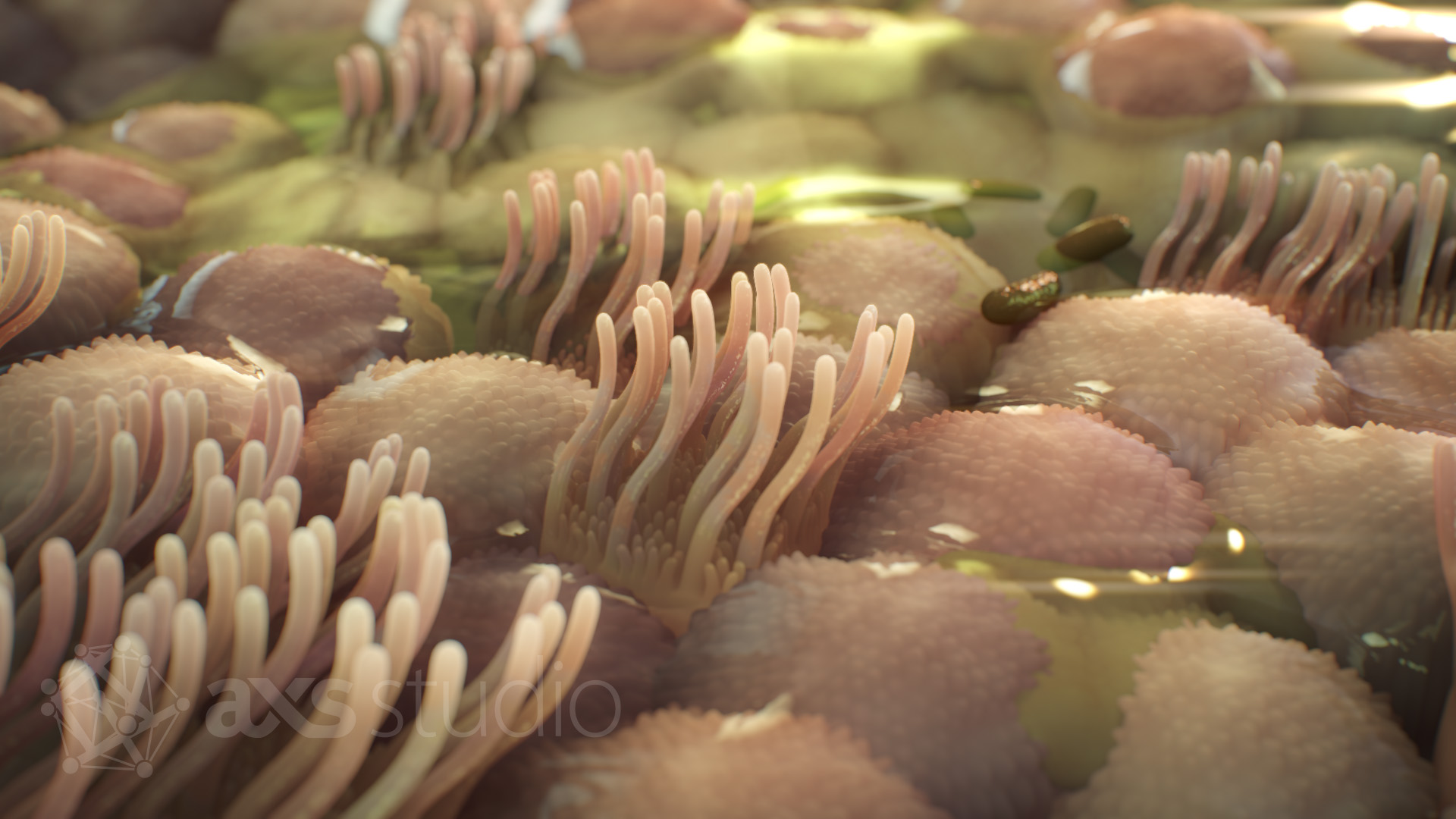
Sea anemones? Nope! These broom-like structures are the cilia that line your respiratory tract. These tiny brooms help sweep mucus and debris up and out of your lungs. This video (starting at 2:06) shows how they work collectively to keep your lungs clean and what happens when they’re unable to function, as is the case with cystic fibrosis.
- Your combined airways stretch from Chicago to Las Vegas. An intricate, branched network of tubes carries oxygen from your mouth/nose to the alveoli, where gas exchange happens. If you were to line up your trachea, bronchi, and bronchioles end-to-end, they would extend 1,500 miles (2,400 kilometers), which is approximately the distance from Chicago to Las Vegas or Toronto to New Orleans.
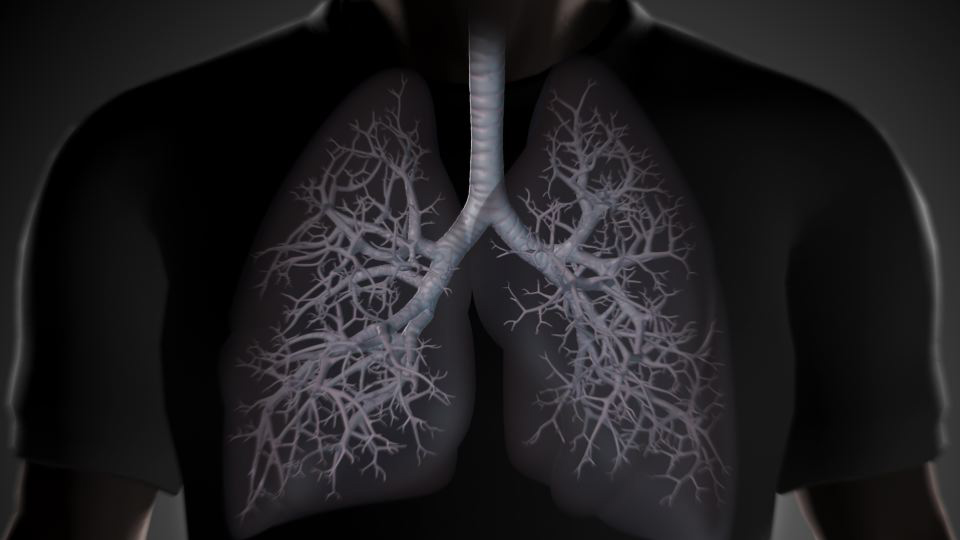
Resembling an upside-down tree, your airway is a highly branched network of tubes that carry oxygen into your body and waste products like carbon dioxide out. The trachea, the trunk of the tree, splits off into large branches called bronchi. The bronchi further split into smaller and smaller branches called bronchioles.
The Liver
Why it’s a vital organ: The ultimate multitasker, your liver is a vital organ that performs over 200 functions. The liver has vital roles in your body, including detoxification, metabolism, and bile production.
Fun facts:
- Your liver is part of your immune system. Your liver has a specialized population of macrophages called Kupffer cells. Only found within liver sinusoids, Kupffer cells engulf pathogens, debris, and foreign materials, removing them from your blood.
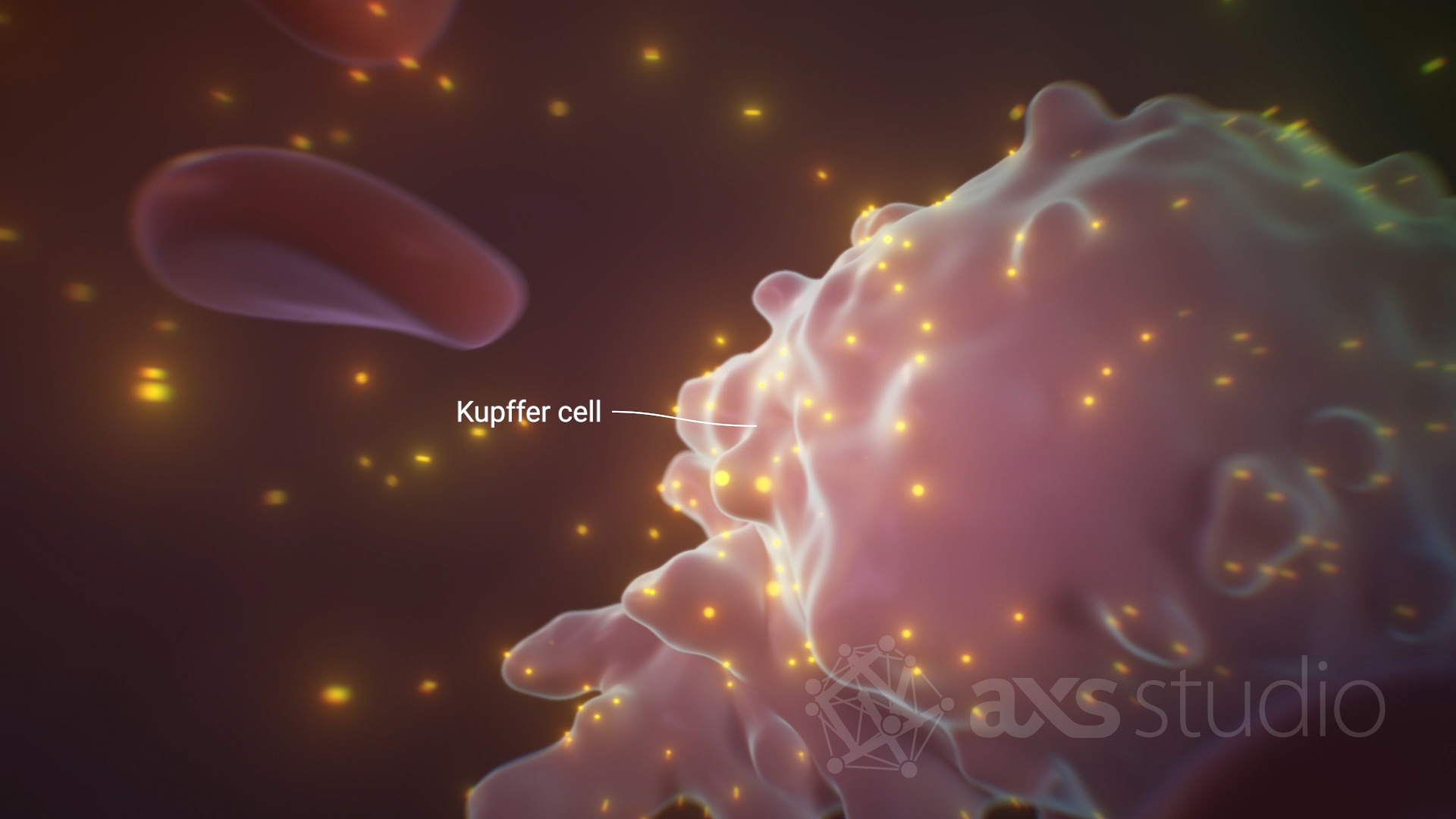
The liver may not be the first organ to come to mind when you think about the immune system. However, immune cells are poised in the liver to mount a quick response to pathogens carried into the liver by the hepatic artery and portal vein. Shown here is a specialized macrophage called a Kupffer cell, sequestering therapeutic nanoparticles from the blood.
- Liver blood vessels are like a spaghetti strainer. Liver sinusoidal endothelial cells, which line the specialized blood vessels of the liver, are fenestrated, meaning they are full of holes. This is a feature, not a defect! The fenestrations facilitate exchange of materials between the blood and cells within the liver.
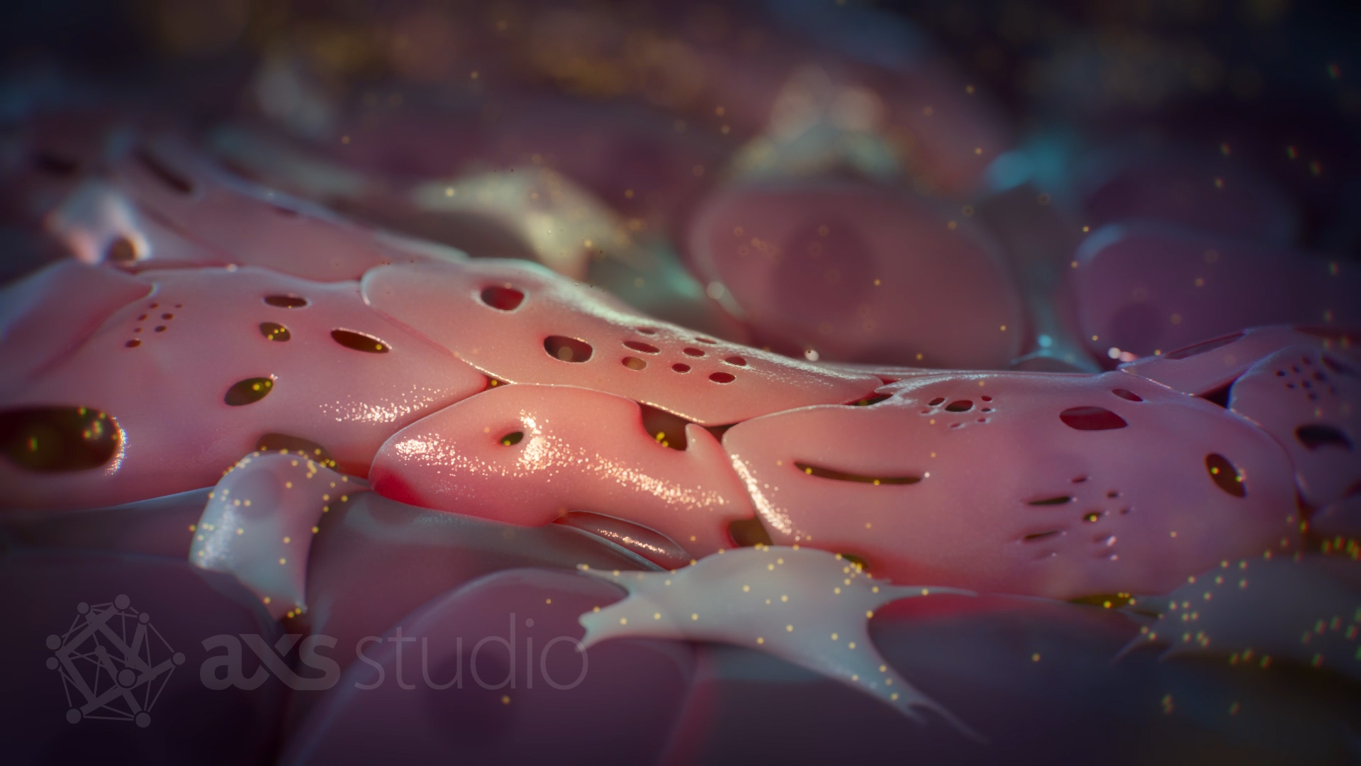
The walls of liver sinusoid blood vessels are very porous and lack a basement membrane. This helps the exchange of materials, like proteins and immune cells, between the bloodstream and liver—the opposite of how the blood brain barrier functions. Here, therapeutic nanoparticles that escaped capture by Kupffer cells leak into the surrounding tissue where they are sequestered by liver cells.
- The Flying Wallenda of blood coagulation. Your liver performs a delicate balancing act in hemostasis by synthesizing both pro- and anti-coagulation factors. Too many pro-coagulation factors, and you risk the formation of a dangerous blood clot. On the other hand, too many anticoagulant proteins, and you’re at risk of excessive bleeding. A healthy liver helps keep things in balance—an amazing feat!
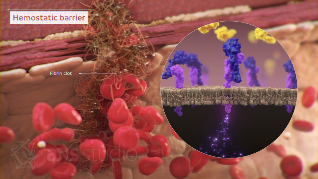
Most of the proteins involved in hemostasis—the regulation of blood flow—are produced by the liver. Blot clots, or thrombi, are essential to stop bleeding at the sites of blood vessel injury. But in the world of hemostasis, sometimes you can have too much of a good thing. Your liver makes sure clotting factors are balanced with anticoagulants, so that vessels are not accidentally blocked by dangerous clots that may cause serious health problems such as heart attack or stroke.
The Kidneys
Why it’s a vital organ: The dynamic duo, your kidneys are vital organs that remove waste from your body, regulate fluid and electrolyte balance, and maintain blood pressure. The kidneys also synthesize hormones involved in stimulating production of red blood cells and the active form of vitamin D.
Fun facts:
- Team work makes the dream work. Each kidney is equipped with a million mini filtration units called nephrons. Nephrons work as a team to remove waste and extra fluid from your blood, which will eventually be excreted by your body as urine. Filtration starts in the glomerulus, where waste products and excess water are filtered out. Next, the filtrate moves through the loop of Henle, where important salts, nutrients, and water can be reabsorbed into the blood stream. This two-step process helps your kidneys maintain a healthy fluid and electrolyte balance.
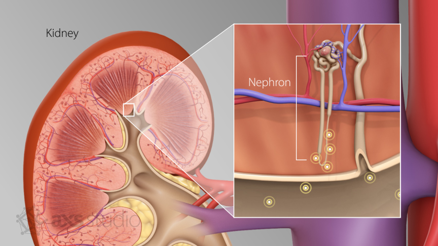
Nephrons are the structures in your kidneys responsible for filtering blood and producing urine. Nephrons have a distinctive structure shown here. Blood enters the nephron through a network of capillaries called the glomerulus—the site where filtration begins. Here, small particles are forced from the blood into a surrounding cup-like structure called Bowman’s capsule. Filtrate moves through the renal tube, including the loop of Henle. There, useful nutrients, solutes, and fluid can be reabsorbed into the bloodstream so they are not accidentally excreted in the urine.
- Not everyone has two kidneys. Although exceedingly rare, some people are born with an extra kidney—called supernumerary kidney. Having an extra kidney is rare and usually doesn’t cause symptoms, so it’s hard to know exactly how many people have the condition. However, it’s estimated that about 0.04% of the world’s population have been walking around with a third kidney from birth. More commonly, a person may have three (or more!) kidneys because they have been the recipient of one or more kidney transplants. In many cases, the recipient’s original kidneys are kept in place, and the new kidney is placed in the lower abdomen. Similarly, there are people out there who have just one kidney. A person may have one kidney from birth or may have had one kidney removed due to disease or live donation. Only one kidney is needed to live a full and healthy life.
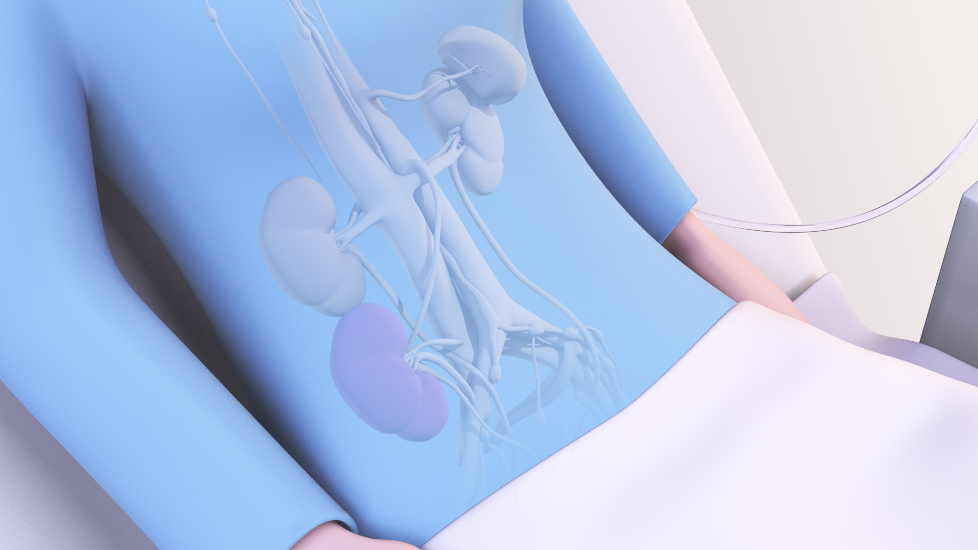
It’s not unusual for transplant recipients to have more than two kidneys. This image from a medical animation shows a patient post-kidney transplant. Her original kidneys were left in their original position in the upper abdomen. The transplanted kidney, shown in purple, is placed in the lower abdomen, close to the bladder and major blood vessels.
FAQ About Vital Body Organs
Vital body organs are those that are essential for survival. In the human body, the vital organs are: the brain, heart, lungs, liver, and kidneys.
Humans have several organs they can live without, albeit medical intervention is usually required. Examples of non-essential organs include the spleen, gallbladder, appendix, and parts of the gastrointestinal tract like the stomach and colon. And despite the lungs and kidneys being vital organs, you actually only need one of each!
Depends on who you ask! The human body has at least 78 organs, with 5 being considered vital organs (brain, heart, lungs, liver, and kidneys). The mesentery—a band of membrane that holds your abdominal organs in place—has recently been crowned organ number 79. And some scientists argue there are actually 80 organs including the interstitium—a network of connective tissue and fluid-filled spaces in and between tissues.
Our vital organs and other body organs work together in groups—or organ systems—to perform bodily functions. Humans have 11 organ systems: respiratory, digestive/excretory, circulatory, urinary, integumentary, skeletal, muscular, endocrine, lymphatic, nervous, and reproductive systems.
Making up a staggering 16% of your total body mass, your skin is the largest organ. The largest internal organ is one of the vital body organs, the liver. The smallest organ is the pineal gland, which is about the size of a grain of rice.
The most common whole-organ transplants are: kidney, liver, heart, pancreas, lung, and small intestine. Your brain is the only vital organ of the body that cannot be replaced by a transplanted organ, so take care of the one you have!
Yes, temporarily. Mechanical artificial hearts can be used to pump blood in people waiting for a heart transplant. Similarly, extracorporeal membrane oxygenation (ECMO) is an external mechanical device that oxygenates blood and removes carbon dioxide, mimicking the function of the lungs. Artificial livers are used to filter blood in patients with acute liver failure who are waiting for a transplant. Artificial kidneys and advancements in other artificial organs are an active area of research and development.
Yes. The liver has the amazing ability to regenerate–it can grow back to full size after up to 90% has been removed. To a lesser extent, the kidneys and heart also regenerate in order to repair minor damage.
References
Ahmed Nada, Ash Jhamb, Julian Maingard (2024). Case Report of Right Supernumerary Kidney in a 38 Year Old Male [article]. Radiol Case Reports 19(9). https://www.sciencedirect.com/science/article/pii/S1930043324005053.
American Lung Association Each Breath Blog (2017, July 19). How Your Lungs Get the Job Done [article]. https://www.lung.org/blog/how-your-lungs-work.
Braet F, Wisse E (2002). Structural and Functional Aspects of Liver Sinusoidal Endothelial Cell Fenestrae: A Review [article]. Comp Hepatol 1(1 ). https://doi.org/10.1186/1476-5926-1-1.
Cleveland Clinical Health Library (2023, November 21) Broken Heart Syndrome [article]. https://my.clevelandclinic.org/health/diseases/17857-broken-heart-syndrome.
Cleveland Clinic Health Library (2024, December 9). Organs in the Body: Definition & Anatomy [article]. https://my.clevelandclinic.org/health/articles/organs-in-the-body.
Columbia University Irving Medical Center (n.d.). Your Heart Has a Brain of Its Own [article]. https://columbiasurgery.org/news/your-heart-has-brain-its-own.
Dotiwala AK, McCausland C, Samra NS (2023, April 4). Anatomy, Head and Neck: Blood Brain Barrier. StatPearls [ebook]. Treasure Island (FL): StatPearls Publishing; 2025. https://www.ncbi.nlm.nih.gov/books/NBK519556/.
InformedHealth.org (2023, April 18). In brief: How do Lungs Work? [article]. Institute for Quality and Efficiency in Health Care (Cologne, Germany); 2006: https://www.ncbi.nlm.nih.gov/books/NBK401240/.
Kalra A, Yetiskul E, Wehrle CJ, et al. (2023, May 1). Physiology, Liver. StatPearls [ebook]. Treasure Island (FL): StatPearls Publishing; 2025. https://www.ncbi.nlm.nih.gov/books/NBK535438/.
Kidney Foundation of Canada (n.d.) How Kidneys Work [webpage]. https://kidney.ca/Kidney-Health/Your-Kidneys/How-Kidneys-Work.
Mayo Clinic (2025, January 31). Kidney Transplant [webpage]. https://www.mayoclinic.org/tests-procedures/kidney-transplant/about/pac-20384777.
Medical Advisory Secretariat (2006, March 1). Ablation for Atrial Fibrillation: An Evidence-based Analysis [article]. https://pmc.ncbi.nlm.nih.gov/articles/PMC3379526/.
National Institute of Diabetes and Digestive and Kidney Disease (2018, June). Your Kidneys & How They Work [webpage]. https://www.niddk.nih.gov/health-information/kidney-disease/kidneys-how-they-work.
Physiopedia (n.d.) Vital Organs [article]. https://www.physio-pedia.com/Vital_Organs.
Ripa R, George T, Shumway KR, et al. (2023, July 30). Physiology, Cardiac Muscle. StatPearls [ebook]. Treasure Island (FL): StatPearls Publishing; 2025. https://www.ncbi.nlm.nih.gov/books/NBK572070/.
Singh R, Munakomi S (2023, May 1). Embryology, Neural Tube. StatPearls [ebook]. Treasure Island (FL): StatPearls Publishing; 2025. https://www.ncbi.nlm.nih.gov/books/NBK542285/.
Thau L, Reddy V, Singh P (2022, October 10). Anatomy, Central Nervous System. StatPearls [ebook]. Treasure Island (FL): StatPearls Publishing; 2025. https://www.ncbi.nlm.nih.gov/books/NBK542179/.
Thrombosis Australia (n.d.). Your liver and the Coagulation Cascade [article]. https://www.pbi.org.au/news/your-liver-and-the-coagulation-cascade.