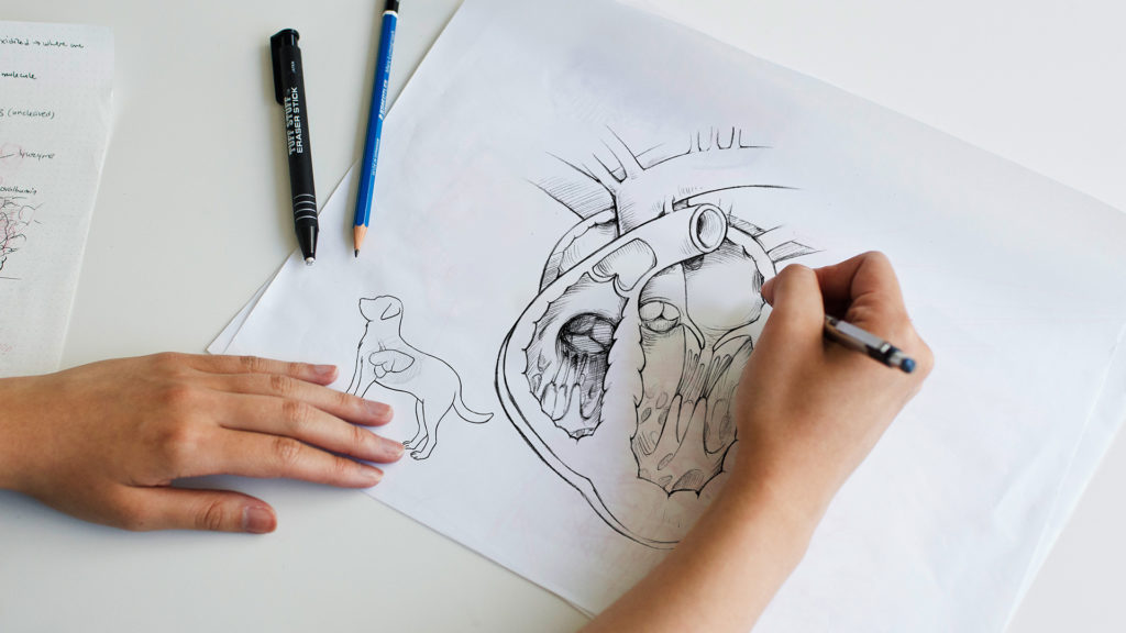
Medical, nursing and allied health students have long relied on the work of medical illustrators to learn the complexities of human anatomy. With the advent of 3D animation technology, a new learning tool became available: 3D medical animation videos. No longer were images confined to two dimensions on the printed page. Educators could now show structures and relationships in three dimensions from different views. And because animation is a temporal medium, it became possible to show continuous multistep processes, of which there are many within the large scope of medical education.
3D Medical Animation Can Visualize Complex Structures and Processes for Medical Students
3D medical animation videos can help learners understand material that is difficult to describe in text, is hidden deep inside the human body, or is even too small to be seen by a microscope. Most medical animation depicts structures at one or more of the following scales: gross anatomical, cellular, and molecular. Medical and nursing students study gross anatomy when learning about body systems and organs, to correctly interpret x-rays, MRIs and CT scans, and to gain an understanding of surgeries and other procedures. They learn about cells and tissues to gain an understanding of physiology, metabolism and pathology. And these future healthcare professionals study molecular biology and biochemistry to learn about the structure of macromolecules in the body, genetics, signalling, the molecular basis of disease, and drug targeting and interaction. When properly trained in accredited graduate-level medical illustration programs, medical animators can visualize these structures and processes at the appropriate level of detail and accuracy for future doctors and nurses.
Medical education covers many physiologic processes like muscle contraction, shown here. 3D animation enables us to show these processes in slow motion with colour-coding to make them easy to follow and understand.
Medical Animation Videos of Gross Anatomy for Education
Anatomical illustration is commonly used in textbooks and atlases to visualize organs and tissues. If you’ve had the experience of looking at photographs of medical specimens, you know that it can be tricky to identify and understand the individual structures. Illustration has the advantage of selective use of colour and contrast to highlight specific elements and can therefore be easier to interpret than photographs. Likewise with animation. The medical animator, like the illustrator, decides when and where to de-emphasize or omit elements that are not part of the story in order to remove distractions and enhance visual clarity. When visualizing intricate organs, tissues or vessels, an animation can distill highly complex structural detail into visuals that are much easier to understand. Animation has advantages over static 2D illustration in that it can show anatomical structures from different views, using depth and perspective to clarify spatial relationships. Animation can also show movement and change over time, which can make it easier to understand processes like blood circulation and muscle contraction, as well as sequential processes like surgical procedures.
The Cellular Scale in 3D Medical Animation Video
Clearly depicting processes at the level of cells is a key element of many medical animations created for medical education. Cells can be seen under a microscope, but it can be difficult to understand a histological section, a blood smear, or other micrographs without extensive experience and knowledge. Using animation, a specific cell type or process can be highlighted in order to help the viewer understand key details. In 3D animation, the animator controls a virtual camera that behaves much like a movie camera, recording events as they unfold within a digital scene. Using the camera to explore a cellular environment, the animator gives viewers a sense of the arrangement and structure of the different cell types in 3D space. Cellular motion in 3D space, such as migration, can also be shown through animation in a way that it is difficult to capture with static illustration or microscopy.
This shot from a medical animation about wound healing depicts fibroblast cells migrating over a network of collagen fibers. We visualized the cytoskeleton within the cells to highlight its involvement in cellular motility. Medical animators model cells using microscope images for reference. They use video microscopy and other peer-reviewed literature to guide their depictions of cell movement.
Getting Tiny with it: 3D Medical Animation at the Scale of Molecules
Visualizing the world at the scale of molecules is a particular challenge. Currently, the most powerful microscopes are able to capture fuzzy images of individual molecules under specific conditions and the ability to capture movement is very limited. In contrast, 3D animation is a powerful tool to visualize molecular structures and processes at high-resolution.
The specific structure of many proteins and nucleic acids (like DNA) are obtained using techniques developed by physicists, including x-ray crystallography and nuclear magnetic resonance. The data obtained, (specifically the exact coordinates in 3D space of each atom in the molecule), are published in public datasets, which medical animators can use to create accurate and detailed 3D molecular models. With these, animators can create didactic visualizations that teach medical students about the structure, motion and interactions of molecules relevant to human health and disease. For example, a 3D animation can show students how an enzyme interacts with its target to catalyze a reaction, or how the 3D structure of a receptor molecule determines its ability to bind a specific ligand.
In this clip from a 3D medical animation, a drug molecule binds tightly within a ligand-binding pocket of a protein receptor. The animation allows a better understanding of the three dimensional physical interactions between the two molecules and highlights the three specific spots that enhance binding affinity and specificity.
Can 3D Animation Video Make Medical Education More Engaging?
An eye-catching medical animation can draw viewers in and hold their attention, making it a useful tool in medical education. Engaged learners retain material better and this is as true for medical, nursing, and allied health students as it is for a patient trying to understand their medical condition. Animation is often coupled with narration to aid in the understanding of unfamiliar visuals. Medical animators also leverage best practices from filmmaking (think Hollywood movies) to focus attention and guide viewers through a story. Cinematography, pacing, shot transitions, lighting and composition can all be used to engage viewers, hold their interest, and draw them into the story while teaching them about complex topics.
Calling David Attenborough: The Narration Script and Voice Actor in 3D Medical Animation
3D medical animation videos invite learners to take in a story with their ears as well as their eyes. While text and static illustration are valuable teaching tools, animation can be even more effective in guiding the viewer through complex material. A well-crafted script voiced by an experienced narrator greatly enhances the learning experience, improving viewers’ ability to understand and retain the material. By crafting the voiceover and visuals to work together, we can focus viewer attention exactly where it needs to be at each moment in the story. When handled correctly, audio and video complement the teaching power of each other. In contrast, if the narration and visuals do not align, it’s a sure way to lose and confuse the audience.
Made You Look: Using the Visual Tools of Animation to Direct Attention
3D medical animation videos can make use of a wide variety of visual tools to direct a viewer’s attention and help them focus on key elements of the story. Even in a scene rich in visual detail, a skilled medical animator is able to make the important “characters” stand out using one or more of the following tools.
Colour and Contrast
Using a constrained colour palette and reserving more saturated colours for the key elements of a story helps viewers quickly recognize what’s important and what isn’t—a concept we call visual hierarchy. Reserving the areas of highest contrast for the focal point of the shot helps direct the viewer’s attention subconsciously.
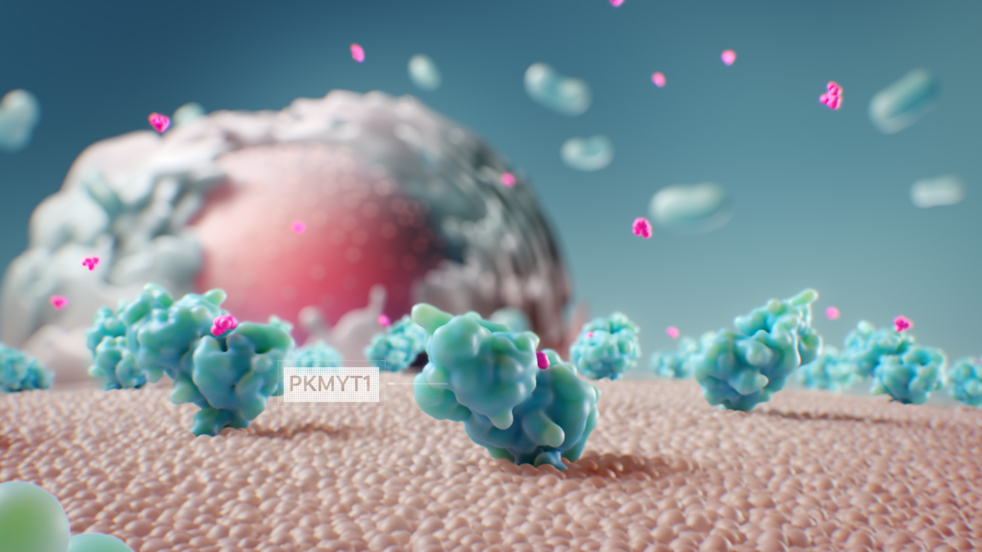
In this frame from a medical animation about a cancer drug, colour is used effectively to guide the viewer’s attention. The colour palette is limited, and complimentary colours—cyan and pink—produce high contrast that draws the eye to important events on screen: the binding of drug molecules to their targets on the surface of a golgi apparatus.
Shot Composition
The layout of elements on a screen can be used to guide the eye to the important details. The animator can position the camera to keep the important characters at the focal point of the composition, and use converging lines to direct the viewer’s attention to that point.
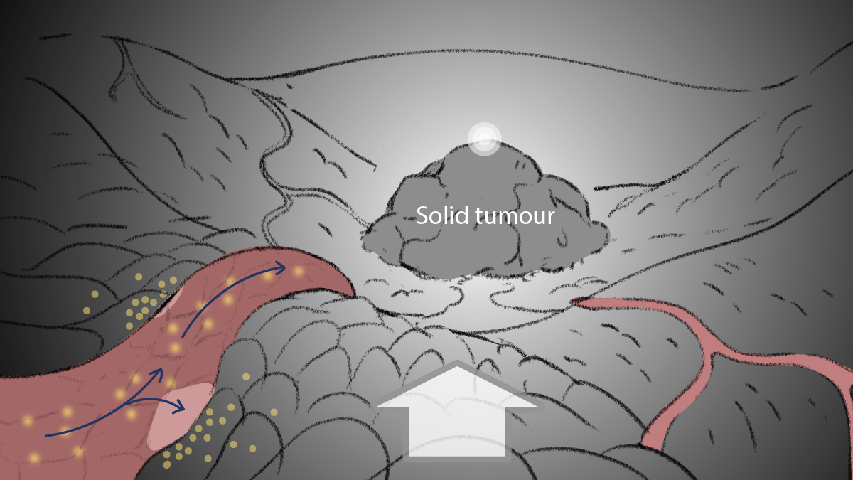
The lines of the blood vessels and the contours of the cellular environment direct attention to the focal point of the image in this storyboard panel from an animation about nanoparticle delivery to cancerous tumours.
Depth of field (DOF)
In the real world, a camera has a focal plane, and objects in the scene become more and more out of focus the further they are in front of or behind the focal plane. In 3D medical animation, animators often add this focal effect, both to emulate the look of real world footage and as a tool to direct the viewer’s attention. The key elements of the shot are kept in sharp focus and visually separated from a background that is blurred. Out-of-focus foreground elements between the camera and the subject can be positioned at the edges of the image to further emphasize the focal point and add to the illusion of depth.
In this frame from a 3D medical animation about SARS-CoV-2 virus detection, DOF is used to direct the audience’s gaze. The objects of interest are kept in sharp focus while the soft background and foreground focus contribute the sense of spatial depth.
Motion
Humans are hard wired to notice motion in our field of view. Medical animators leverage this characteristic to subtly “tell” viewers where to look in a scene. If most of the objects in the frame are static or moving slowly, the viewer’s attention can be drawn to elements moving more quickly. Motion graphics—animated text and graphical elements—work by drawing a learner’s attention to elements in a specific order, using movement on screen. Medical animators often employ motion graphics for labels and to describe complex biological processes in more simple terms.
Icons draw attention as they animate in, and are then highlighted in sync with the voiceover to add emphasis and make the story easier to follow.
Camera Movement
The medical animator has a wide variety of camera moves at their disposal to help direct and hold the viewer’s attention, and add interest and clarity to the storytelling. For example, the camera can push in on a group of cells to highlight an event. Or the camera might pull back from a small tissue environment to reveal where it’s located in the body. In a tracking shot, the camera follows a character as they move through their environment which can give a sense of their journey. A slow and smooth camera can evoke calm, while quicker camera moves can give a sense of urgency or tension. When used skillfully by an experienced animator, camera moves can make 3D medical animation more engaging for a medical education audience.
This 3D animation clip is set inside the cytoplasm of a cell. In it, the camera travels through a mass of misfolded proteins above the endoplasmic reticulum to “find” a proteasome, whose job it is to break down and recycle these problematic proteins within cells. In this way, the camera helps tell the story in a few short seconds: first showing us the problem, then arriving at the solution.
Frequently Asked Questions
3D medical animation is created by specially trained medical animators. It employs specialized knowledge and 3D animation software to visualize anatomical structures, physiological and molecular processes, and medical procedures. It is used in medical education to help teach complex concepts by providing dynamic and engaging visual content.
Compared to traditional methods, evidence suggests that 3D animation can enhance engagement, comprehension and retention. It can do this by providing spatial understanding of structures that might be difficult to understand with text descriptions and 2D static images, and by visualizing dynamic processes and change over time.
Effectiveness can be assessed by testing knowledge gain using pre- and post-tests. Student feedback has been collected to assess usability and appeal. Engagement metrics like view time and repeat views can be used to measure how effectively the animation engages the viewers.
3D medical animation is effective for a broad range of topics, including:
- Anatomy (e.g. an animation teaching the spatial relationships of different regions of the brain)
- Physiology (e.g. an animation depicting the changes in the electrochemical gradient across a cell membrane during neurotransmission)
- Pathology (e.g. a mechanism of disease animation showing progression of cancer, cystic fibrosis or osteoporosis)
- Surgical procedures (e.g. an animation showing the complex steps involved in laparoscopic hernia repair)
- Pharmacological mechanisms (e.g. a mechanism of action animation showing how a drug molecule specifically binds and inactivates its target molecule)
High-quality 3D medical animations are created by specially trained medical animators who use peer-reviewed data, journal articles, medical textbooks, anatomy atlases, micrographs, and 3D molecular data. They are often created in collaboration with medical experts, educators and clinicians.
3D medical animations are made by medical animators specially trained at one of five accredited postgraduate programs. The animations are commissioned by medical education publishers—often the same companies that publish textbooks—and by medical schools and professors. Medical education for patients is commissioned by disease advocacy groups and pharmaceutical companies.
Yes, they can be adapted for use by different audiences. An animation for medical students will include more visual detail and technical information, often with extensive labelling. An animation for patient education will use more accessible language and a more patient-friendly visual style, generally with less detail.
Additional Resources for 3D Medical Animation Videos in Medical Education
- Is Video-Based Education an Effective Method in Surgical Education? A Systematic Review (link)
- Molecular Illustration in Research and Education: Past, Present, and Future (link)
- The use of 3D video in medical education: A scoping review (link)
- Using 3D Animation in Biology Education : Examining the Effects of Visual Complexity in the Representation of Dynamic Molecular Events (link)
One Response
Yes I need to join I am studying in post graduate nursing in jipmer pondicherry I